Tag: orthopedic hospital New BEL Road

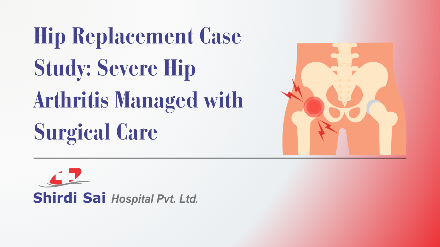
  • Hip Replacement Case Study: Severe Hip Arthritis Managed with Surgical Care

    Hip Replacement Case Study: Severe Hip Arthritis Managed with Surgical Care

    Reviewed by Shirdi Sai Hospital Team

    Severe hip arthritis can make walking, climbing stairs, sitting, standing, and routine daily activities difficult. When hip pain continues for years and does not improve with conservative care, total hip replacement may be considered after orthopedic evaluation. In some complex cases, additional surgical care may be required if a fracture occurs around the hip implant.

    This case study explains how an elderly patient with severe left hip osteoarthritis was evaluated and treated at Shirdi Sai Hospital, New BEL Road. The patient underwent left total hip replacement and later required fracture fixation for a periprosthetic femur fracture. The case is shared for awareness only. Patient identity and personal details have been fully removed for privacy.

    Case Summary

    An elderly female patient came to Shirdi Sai Hospital with severe left hip pain and difficulty walking. The pain had been present for several years and had worsened over time. The patient was unable to walk comfortably, climb stairs, or perform routine activities without significant difficulty.

    The patient had already been evaluated for bilateral hip osteoarthritis and had previously undergone right hip replacement. This admission was for management of severe left hip osteoarthritis.

    After evaluation by the Orthopedics team, the patient underwent left total hip replacement. A post-operative X-ray later showed a periprosthetic fracture of the left femur. The patient then underwent open reduction and internal fixation using a proximal femur plate and stainless steel wire. The post-operative course was stable, and the patient was discharged with medicines, physiotherapy advice, walking guidance, catheter removal advice, and follow-up instructions.

    What Was the Patient’s Main Problem?

    The patient’s main problem was long-standing severe pain in the left hip. The pain affected basic movement and daily activity.

    The patient had difficulty with:

    • walking
    • climbing stairs
    • doing routine activities
    • standing or moving comfortably
    • maintaining normal mobility

    Long-standing hip pain can reduce independence, confidence, and quality of life, especially in elderly patients. When hip arthritis becomes advanced, patients may avoid walking due to pain, which can further reduce muscle strength and mobility.

    Final Diagnosis

    The final diagnosis included:

    • left hip osteoarthritis
    • left femur periprosthetic fracture
    • previous right hip replacement in situ

    This means the patient had severe arthritis of the left hip and later developed a fracture around the femur near the hip replacement implant. The right hip replacement had already been done earlier and was present at the time of admission.

    What Is Hip Osteoarthritis?

    Hip osteoarthritis is a degenerative joint condition where the cartilage covering the hip joint gradually wears down. As cartilage reduces, the bones may rub against each other, causing pain, stiffness, reduced movement, and difficulty walking.

    Common symptoms of hip osteoarthritis include:

    • groin pain
    • hip pain
    • thigh pain
    • stiffness after rest
    • difficulty walking
    • difficulty climbing stairs
    • difficulty sitting cross-legged
    • pain while standing from a chair
    • reduced hip movement
    • limp while walking
    • sleep disturbance due to pain in some patients

    Hip osteoarthritis usually progresses gradually. In early stages, medicines, exercise, weight control, walking support, and physiotherapy may help. In advanced stages, when pain and disability become severe, surgical treatment may be considered.

    Patients with hip pain, knee pain, fractures, arthritis, sports injuries, or mobility problems can consult the Orthopedics department at Shirdi Sai Hospital for evaluation.

    When Is Hip Replacement Considered?

    Hip replacement is considered when arthritis has severely damaged the hip joint and symptoms are affecting daily life despite conservative care.

    It may be discussed when the patient has:

    • severe hip pain
    • difficulty walking
    • difficulty climbing stairs
    • reduced range of hip movement
    • pain not improving with medicines or physiotherapy
    • X-ray evidence of advanced hip arthritis
    • inability to perform routine activities
    • poor quality of life due to hip pain

    Hip replacement is not advised only because an X-ray shows arthritis. The decision is based on symptoms, functional limitation, imaging findings, age, medical condition, and patient-specific goals.

    Treatment Approach at Shirdi Sai Hospital

    The patient was admitted under orthopedic care. The treatment process included:

    • orthopedic evaluation
    • clinical examination
    • relevant investigations
    • review of reports
    • surgical consent
    • left total hip replacement
    • post-operative monitoring
    • post-operative X-ray assessment
    • blood transfusion support when required
    • second surgery for fracture fixation
    • antibiotics and supportive medicines
    • pain management
    • physiotherapy
    • walker-assisted mobilisation
    • discharge planning
    • follow-up advice

    The care plan was based on the patient’s condition, diagnosis, operative findings, and recovery status.

    What Is Total Hip Replacement?

    Total hip replacement, also called total hip arthroplasty, is a surgery where the damaged parts of the hip joint are replaced with artificial components. It is commonly done for advanced hip arthritis when pain and stiffness affect mobility.

    The goal of hip replacement is to:

    • reduce arthritis-related hip pain
    • improve hip function
    • help the patient walk better
    • improve daily activity ability
    • support better mobility when recovery is complete

    Recovery after hip replacement requires physiotherapy, walking support, wound care, fall prevention, and follow-up visits.

    What Is a Hybrid Hip Replacement?

    A hybrid hip replacement is a type of hip replacement where the surgeon uses a combination of fixation methods for the implant components. The exact implant choice depends on bone quality, patient age, surgeon assessment, and intraoperative decision-making.

    In this case, the patient underwent left total hip replacement using a hybrid approach. The choice of technique was made by the treating orthopedic team based on clinical judgment.

    What Is a Periprosthetic Femur Fracture?

    A periprosthetic femur fracture is a fracture that occurs around or near a hip replacement implant. It is a known orthopedic complication and needs careful evaluation because treatment depends on the fracture location, implant stability, bone quality, and patient condition.

    A periprosthetic fracture may require:

    • observation in selected stable cases
    • fixation using plate, screws, cables, or wires
    • revision hip surgery in selected cases
    • restricted or guided weight bearing
    • physiotherapy
    • close follow-up

    In this case, the post-operative X-ray showed a left femur periprosthetic fracture. The patient underwent open reduction and internal fixation with a proximal femur plate and stainless steel wire.

    What Is ORIF with Plate and Wire?

    ORIF means Open Reduction and Internal Fixation.

    • Open reduction means the fracture is surgically exposed and aligned.
    • Internal fixation means implants are used to hold the bone in position while it heals.

    In this case, the fracture was fixed using a proximal femur plate and stainless steel wire. This helped stabilize the fracture around the hip replacement area.

    ORIF is planned based on fracture pattern, bone quality, implant position, and patient safety.

    Why Was a Second Surgery Needed?

    A second surgery was needed because a periprosthetic fracture was identified after hip replacement. Such fractures need proper stabilization when the fracture pattern and clinical condition require it.

    The goal of the second surgery was to:

    • align the fractured femur
    • stabilize the bone
    • protect the hip replacement area
    • support recovery and mobility planning
    • reduce the risk of poor fracture healing

    The patient underwent ORIF with proximal femur plate and stainless steel wire. The operative and post-operative course was documented as stable.

    Hospital Course and Recovery

    After the hip replacement surgery, the patient was shifted for monitoring. Post-operative orders were followed, vitals were monitored, and imaging was reviewed.

    After the periprosthetic fracture was identified, the patient underwent fracture fixation surgery. The patient was later shifted for ward observation once stable.

    The recovery plan included:

    • post-operative monitoring
    • antibiotics and supportive medicines
    • pain control
    • blood transfusion support when required
    • wound dressing
    • physiotherapy
    • walker-assisted mobilisation
    • discharge medicines
    • follow-up review
    • catheter removal advice

    At discharge, the patient was documented as hemodynamically stable.

    Physiotherapy After Hip Replacement and Fracture Fixation

    Physiotherapy is a key part of recovery after hip replacement and fracture fixation. Surgery stabilizes the joint and bone, but movement, strength, balance, and walking confidence need guided rehabilitation.

    Physiotherapy may help with:

    • safe movement training
    • strengthening exercises
    • walking with support
    • fall prevention
    • reducing stiffness
    • improving confidence
    • protecting the operated limb
    • gradual return to daily activities

    Patients should follow the treating doctor and physiotherapist’s instructions about walking, weight-bearing, exercises, and movement restrictions. These instructions can differ from patient to patient.

    Warning Signs After Hip Replacement or Fracture Fixation

    The discharge advice included immediate reporting if persistent fever or pus at the operated site occurs.

    Patients should seek urgent review if they notice:

    • persistent fever
    • pus discharge from wound
    • wound redness
    • wound swelling
    • severe pain
    • increasing hip or thigh swelling
    • bleeding from wound
    • calf pain or swelling
    • breathing difficulty
    • sudden inability to move the operated limb
    • fall after surgery
    • sudden worsening of pain
    • catheter-related discomfort, blockage, or reduced urine output

    Post-operative symptoms should not be ignored, especially in elderly patients.

    What This Case Teaches Patients About Hip Arthritis

    This case highlights an important lesson: long-standing hip pain should not be ignored when it starts affecting walking, stairs, and daily routine.

    Key learnings include:

    • Severe hip arthritis can affect independence.
    • Difficulty walking and climbing stairs needs orthopedic evaluation.
    • Hip replacement may be considered in advanced arthritis when symptoms are severe.
    • Periprosthetic fracture is a serious complication that needs specialist management.
    • Recovery after hip surgery needs physiotherapy and follow-up.
    • Elderly patients need careful post-operative monitoring.
    • Treatment decisions vary from patient to patient.

    Hip Arthritis vs Normal Age-Related Hip Pain

    Symptom PatternMore Likely Age-Related StiffnessMore Concerning for Hip Arthritis
    Mild stiffness after restCommonCan occur
    Severe pain while walkingLess typicalCommon in advanced cases
    Difficulty climbing stairsMay occur mildlyCommon when arthritis progresses
    Pain for yearsNeeds evaluationStrong concern
    Reduced hip movementLess typicalCommon
    Limp while walkingNot normalNeeds evaluation
    Inability to do routine workNot normalNeeds orthopedic care
    X-ray showing joint damageNot assessed clinicallySupports diagnosis when symptoms match

    This table is for awareness only. Diagnosis requires clinical examination and imaging.

    Shirdi Sai Hospital and Sathya Sai Hospital Are Different

    Some patients searching online may confuse Shirdi Sai Hospital with Sathya Sai Hospital because the names sound similar. They are not the same hospital.

    This case study refers to Shirdi Sai Hospital Pvt. Ltd., located at No. 519, 2nd Main, Nethravathi Street, Devasandra, New BEL Road, Bangalore – 560054.

    Before visiting, patients should confirm the hospital name, location, phone number, and official website. For this case study and related orthopedic care, the correct hospital is Shirdi Sai Hospital on New BEL Road.

    Patients can visit the official Shirdi Sai Hospital website for hospital information and appointments.

    Why Patients Choose Shirdi Sai Hospital for Hip and Fracture Care

    At Shirdi Sai Hospital, New BEL Road, orthopedic symptoms such as hip pain, knee pain, fractures, arthritis, sports injuries, and mobility problems are evaluated through the Orthopedics department. In complex cases, a multispeciality hospital setting can also support medical evaluation, post-operative monitoring, transfusion support when required, physiotherapy, and follow-up care.

    Patients may choose Shirdi Sai Hospital because:

    • orthopedic care is available on New BEL Road
    • hip arthritis and fracture-related symptoms are evaluated clinically
    • imaging and surgical planning are guided by orthopedic assessment
    • hip replacement and fracture fixation may be considered where clinically suitable
    • post-operative monitoring and physiotherapy guidance support recovery
    • emergency and multispeciality support are available when needed
    • the hospital is accessible for patients from Devasandra, Mathikere, RMV 2nd Stage, Sanjaynagar, Hebbal, and nearby areas

    Patients with severe hip pain, difficulty walking, suspected fracture, arthritis symptoms, or previous joint replacement concerns can consult the Orthopedics department at Shirdi Sai Hospital for evaluation.

    Patient Privacy Note

    This case study has been written with patient identity removed. Personal identifiers such as name, registration number, address, exact admission date, discharge date, and identifying personal details have not been included.

    The purpose of this case study is patient education and awareness. Treatment decisions vary from patient to patient. A similar diagnosis does not mean every patient will need the same surgery, medicines, hospital stay, walking plan, catheter support, or rehabilitation protocol.

    Frequently Asked Questions

    What is hip osteoarthritis?

    Hip osteoarthritis is a degenerative condition where the cartilage in the hip joint wears down, leading to pain, stiffness, reduced movement, difficulty walking, and trouble climbing stairs.

    When is hip replacement needed?

    Hip replacement may be considered when hip arthritis causes severe pain, walking difficulty, reduced movement, and poor daily function despite non-surgical treatment.

    What is a periprosthetic femur fracture?

    A periprosthetic femur fracture is a fracture that occurs around or near a hip replacement implant. It needs orthopedic evaluation because treatment depends on fracture pattern and implant stability.

    What is ORIF in fracture treatment?

    ORIF means Open Reduction and Internal Fixation. It is a surgery where the fracture is aligned and stabilized using implants such as plates, screws, rods, wires, or other fixation devices.

    Is physiotherapy needed after hip replacement?

    Yes. Physiotherapy helps with safe walking, strengthening, movement recovery, stiffness reduction, and fall prevention after hip replacement.

    What warning signs need review after hip surgery?

    Persistent fever, pus discharge, wound redness, severe pain, sudden swelling, breathing difficulty, fall after surgery, or sudden worsening of movement should be reviewed urgently.

    Is every hip arthritis patient treated with surgery?

    No. Treatment depends on severity, pain level, X-ray findings, function, age, health condition, and response to non-surgical care. Surgery is considered when symptoms are advanced and disabling.

    Conclusion

    This case shows how severe left hip osteoarthritis affecting walking, stair climbing, and routine activity was treated surgically at Shirdi Sai Hospital, New BEL Road. The patient underwent left total hip replacement and later required ORIF with proximal femur plate and stainless steel wire for a periprosthetic femur fracture. The patient was monitored, supported with medicines, mobilized with guidance, and discharged in stable condition with follow-up advice.

    Long-standing hip pain should not be ignored when it starts affecting daily life. Early orthopedic evaluation helps patients understand whether medicines, physiotherapy, walking support, hip replacement, fracture fixation, or another treatment plan is suitable.

    OPD Consultation starts from ₹500.
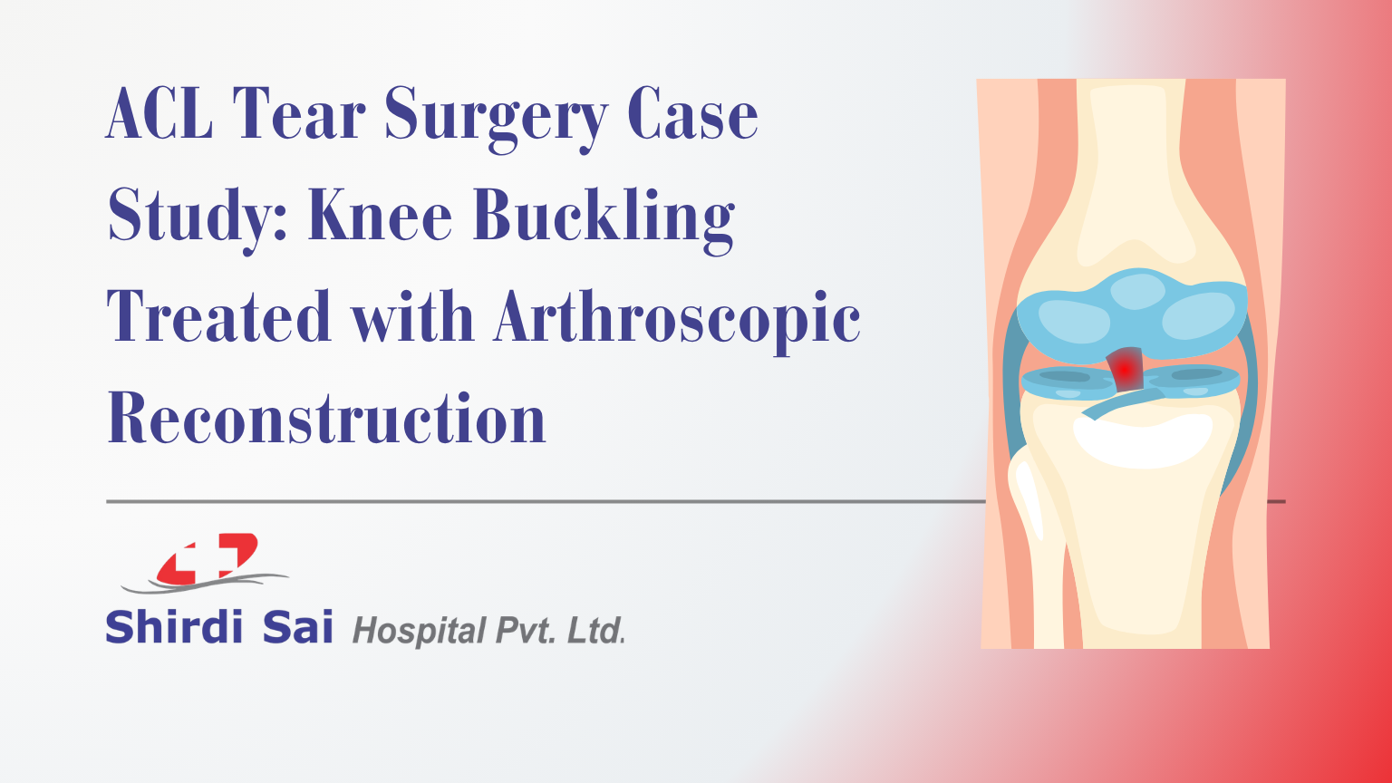
  • ACL Tear Surgery Case Study: Knee Buckling Treated with Arthroscopic Reconstruction

    ACL Tear Surgery Case Study: Knee Buckling Treated with Arthroscopic Reconstruction

    Reviewed by Shirdi Sai Hospital Team

    An ACL tear can cause knee instability, pain, buckling, and difficulty walking confidently. In some patients, the injury may begin after a twisting movement and continue for months if the ligament does not heal properly or if another knee structure, such as the meniscus, is also injured. When knee buckling increases and daily movement becomes difficult, orthopedic evaluation becomes important.

    This case study explains how an adult patient with a right knee ACL tear and medial meniscus tear was evaluated and treated at Shirdi Sai Hospital, New BEL Road. The patient underwent arthroscopic ACL reconstruction with partial medial meniscectomy and was discharged in stable condition with physiotherapy and follow-up advice. The case is shared for awareness only, with patient identity fully removed.

    Case Summary

    An adult male patient came to Shirdi Sai Hospital with a history of twisting injury to the right knee several months earlier. The patient had already taken treatment, but symptoms did not settle completely. Over time, pain increased and the right knee started buckling while walking.

    The patient was evaluated by the Orthopedics team and admitted for further management. Based on clinical findings and diagnosis, the patient was treated for:

    • right knee ACL tear
    • medial meniscus tear

    The surgical procedure included:

    • arthroscopic right ACL reconstruction
    • partial meniscectomy of the medial meniscus

    After surgery, the patient was monitored in the hospital. The post-operative course was stable. Pain gradually reduced, oral intake was tolerated, and the patient was able to ambulate with support. The patient was discharged in a hemodynamically stable condition with medicines, brace support, physiotherapy advice, and follow-up instructions.

    What Was the Patient’s Main Problem?

    The main concern was right knee pain with buckling while walking. Knee buckling means the knee suddenly feels unstable, gives way, or feels unable to support body weight properly during movement.

    In this case, the problem began after a twisting injury. This type of injury can damage knee ligaments, meniscus, or other supporting structures. When the ACL is torn, the knee may lose stability, especially during walking, turning, climbing stairs, or sudden direction changes.

    The patient’s symptoms included:

    • previous twisting injury to the right knee
    • persistent pain despite earlier treatment
    • increased pain over recent weeks
    • buckling of the right knee while walking
    • difficulty trusting the knee during movement

    These symptoms suggested that the knee instability needed orthopedic evaluation and treatment planning.

    Final Diagnosis

    The final diagnosis was:

    Right ACL Tear with Medial Meniscus Tear

    The ACL, or anterior cruciate ligament, is one of the main stabilizing ligaments of the knee. It helps control forward movement and rotation of the shin bone in relation to the thigh bone.

    The medial meniscus is a C-shaped cartilage cushion on the inner side of the knee. It helps absorb shock, distribute load, and support smooth knee movement.

    When both the ACL and medial meniscus are injured, patients may experience pain, instability, swelling, catching, locking, or buckling depending on the tear pattern and severity.

    What Is an ACL Tear?

    An ACL tear is an injury to the anterior cruciate ligament inside the knee. It commonly occurs due to twisting, sudden stopping, pivoting, sports injury, fall, or accident.

    Common symptoms of an ACL tear may include:

    • knee pain after injury
    • swelling
    • instability
    • buckling or giving way
    • difficulty walking confidently
    • difficulty turning or changing direction
    • reduced confidence in the knee
    • pain during activity

    Some ACL injuries improve with rehabilitation in selected patients. However, when instability continues, when the knee buckles during daily activity, or when associated meniscus injury is present, surgical treatment may be considered after orthopedic evaluation.

    What Is a Medial Meniscus Tear?

    A medial meniscus tear affects the cartilage cushion on the inner side of the knee. It can occur along with ligament injuries, especially after twisting movements.

    Symptoms may include:

    • pain on the inner side of the knee
    • swelling
    • catching sensation
    • locking sensation
    • pain while bending the knee
    • discomfort while walking or squatting
    • instability when combined with ligament injury

    Treatment depends on the type, location, and severity of the tear. Some tears may be managed with physiotherapy and medicines. Some tears may require arthroscopic treatment, especially if they are causing mechanical symptoms or are associated with other knee injuries.

    Why Was Surgery Considered in This Case?

    Surgery was considered because the patient had a diagnosed ACL tear with medial meniscus tear and ongoing knee buckling while walking. The symptoms had continued despite earlier treatment and were affecting knee stability.

    The decision for surgery was based on:

    • history of twisting knee injury
    • persistent pain
    • increased buckling of the right knee
    • confirmed ACL tear
    • associated medial meniscus tear
    • orthopedic evaluation
    • need to improve knee stability and function

    The goal was not only pain relief. The main goal was to restore knee stability, treat the meniscus injury appropriately, and support safer movement with rehabilitation.

    Patients with similar symptoms can consult the Orthopedics department at Shirdi Sai Hospital for evaluation of knee pain, ACL injury, meniscus tear, sports injury, fracture care, arthroscopy, and rehabilitation guidance.

    What Is Arthroscopic ACL Reconstruction?

    Arthroscopic ACL reconstruction is a minimally invasive knee surgery performed using a small camera called an arthroscope and specialized instruments. The damaged ACL is reconstructed using a graft to help restore knee stability.

    Arthroscopy allows the surgeon to view the inside of the knee joint through small incisions. This approach is commonly used for ligament reconstruction and meniscus-related procedures when surgery is required.

    In this case, arthroscopic right ACL reconstruction was performed as part of the treatment plan.

    What Is Partial Medial Meniscectomy?

    Partial medial meniscectomy means removing only the damaged portion of the medial meniscus while preserving as much healthy meniscus tissue as possible.

    This may be done when the torn part of the meniscus is not suitable for repair or is causing symptoms. The decision depends on tear type, location, tissue quality, stability, and surgeon assessment.

    In this case, partial meniscectomy of the medial meniscus was performed along with ACL reconstruction.

    Treatment Approach at Shirdi Sai Hospital

    The patient was admitted under orthopedic care and evaluated before surgery. The treatment pathway included:

    • orthopedic evaluation
    • pre-operative assessment
    • physician fitness before surgery
    • surgical planning
    • arthroscopic ACL reconstruction
    • partial medial meniscectomy
    • post-operative monitoring
    • antibiotics and supportive medicines as required
    • pain control
    • gradual diet restart
    • mobilization support
    • discharge advice
    • physiotherapy guidance
    • OPD follow-up recommendation

    The post-operative course was documented as uneventful. Pain gradually reduced, and the patient tolerated oral soft diet. The patient was able to ambulate and was discharged in stable condition.

    Recovery Plan After ACL Reconstruction and Meniscus Surgery

    Recovery after ACL reconstruction and meniscus surgery depends on the injury, procedure, patient condition, physiotherapy compliance, muscle strength, and follow-up care.

    In this case, discharge advice included:

    • mobilization with support
    • long knee brace
    • continued physiotherapy
    • straight leg raise exercises
    • isometric quadriceps exercises
    • follow-up review in OPD

    Physiotherapy is a key part of recovery because surgery alone does not restore full strength, balance, and confidence. The knee needs structured rehabilitation to regain movement, muscle control, and stability.

    Why Physiotherapy Matters After ACL Surgery

    After ACL reconstruction, the thigh muscles, especially the quadriceps, can become weak. Swelling, pain, reduced movement, and fear of using the knee can slow recovery.

    Physiotherapy helps with:

    • reducing stiffness
    • restoring knee movement
    • improving quadriceps strength
    • improving walking pattern
    • reducing swelling
    • improving balance
    • supporting gradual return to activity
    • protecting the reconstructed ligament during healing

    Exercises such as straight leg raises and isometric quadriceps exercises are commonly used in early rehabilitation when advised by the treating team.

    Patients should not restart sports, running, heavy lifting, squatting, or twisting activities without orthopedic and physiotherapy clearance.

    ACL Tear with Meniscus Tear: Why Early Evaluation Matters

    An ACL tear with meniscus injury can affect knee function more than an isolated minor knee sprain. If instability continues, the knee may buckle repeatedly, which can increase the risk of further injury.

    Patients should not ignore:

    • knee buckling while walking
    • repeated giving way sensation
    • swelling after activity
    • pain after twisting injury
    • knee locking or catching
    • difficulty climbing stairs
    • difficulty turning quickly
    • inability to trust the knee
    • pain that continues for weeks after injury

    Early diagnosis helps decide whether physiotherapy, bracing, activity modification, arthroscopy, ACL reconstruction, meniscus repair, partial meniscectomy, or another treatment option is suitable.

    Warning Signs After Knee Surgery

    Patients should follow discharge instructions and return for review if warning signs appear.

    Important warning signs include:

    • fever
    • severe pain
    • pus discharge from wound
    • increasing swelling
    • redness around wound
    • wound soakage
    • calf pain or swelling
    • breathing difficulty
    • sudden worsening of knee pain
    • inability to move the limb as advised
    • fall or fresh injury after surgery

    The discharge instructions in this case specifically advised review if fever, severe pain, or wound discharge occurred.

    What This Case Teaches Patients

    This case highlights a common problem: knee ligament injuries are often underestimated when pain temporarily improves. A patient may continue walking after injury, but instability may become clear later as buckling, giving way, or reduced confidence in the knee.

    Key lessons:

    • Twisting knee injuries should not be ignored if pain persists.
    • Knee buckling is an important instability symptom.
    • ACL tears may need surgery when instability affects daily activity.
    • Meniscus tears can occur along with ACL injury.
    • Arthroscopy can be used to treat selected ACL and meniscus injuries.
    • Physiotherapy is essential after surgery.
    • Follow-up care is important for safe recovery.

    Shirdi Sai Hospital and Sathya Sai Hospital Are Different

    Some patients searching online may confuse Shirdi Sai Hospital with Sathya Sai Hospital because the names sound similar. They are not the same hospital.

    This case study refers to Shirdi Sai Hospital Pvt. Ltd., located at No. 519, 2nd Main, Nethravathi Street, Devasandra, New BEL Road, Bangalore – 560054.

    Before visiting, patients should confirm the hospital name, location, phone number, and official website. For this case study and related orthopedic care, the correct hospital is Shirdi Sai Hospital on New BEL Road.

    You can visit the official Shirdi Sai Hospital website for hospital information and appointments.

    Why Patients Choose Shirdi Sai Hospital for ACL and Meniscus Injury Care

    At Shirdi Sai Hospital, New BEL Road, knee injuries are evaluated through the Orthopedics department with attention to symptoms, injury history, instability, pain pattern, imaging, and functional difficulty. The hospital’s Orthopedics services include care for ACL tears, meniscus tears, sports injuries, arthroscopy, fracture care, rehabilitation support, and musculoskeletal conditions.

    Patients may choose Shirdi Sai Hospital because:

    • orthopedic care is available on New BEL Road
    • ACL and meniscus injuries are evaluated clinically
    • arthroscopy is available for selected joint problems
    • surgical and non-surgical options are considered based on the case
    • rehabilitation guidance supports recovery after surgery
    • emergency and multispeciality support are available when needed
    • the hospital is accessible for patients from Devasandra, Mathikere, RMV 2nd Stage, Sanjaynagar, Hebbal, and nearby areas

    For patients with knee buckling, ligament injury, meniscus tear symptoms, or sports-related knee pain, orthopedic consultation at Shirdi Sai Hospital can help identify the next step.

    Patient Privacy Note

    This case study has been written with patient identity removed. Personal identifiers such as name, registration number, address, exact admission date, discharge date, and identifying personal details have not been included.

    The purpose of this case study is patient education and awareness. Treatment decisions vary from patient to patient. A similar diagnosis does not mean every patient will need the same surgery, medicines, hospital stay, brace, or rehabilitation plan.

    Frequently Asked Questions

    What is an ACL tear?

    An ACL tear is an injury to the anterior cruciate ligament, one of the main stabilizing ligaments inside the knee. It can cause pain, swelling, instability, and knee buckling.

    What are the symptoms of ACL tear?

    Common symptoms include knee pain, swelling, instability, buckling, difficulty walking, and loss of confidence while turning, climbing stairs, or changing direction.

    When does an ACL tear need surgery?

    ACL surgery may be considered when the knee remains unstable, buckles during daily activity, or when there is an associated injury such as a meniscus tear. The decision depends on orthopedic evaluation.

    What is arthroscopic ACL reconstruction?

    Arthroscopic ACL reconstruction is a minimally invasive procedure where the damaged ACL is reconstructed using a graft through small incisions and camera-guided instruments.

    What is partial meniscectomy?

    Partial meniscectomy means removing only the damaged portion of the meniscus while preserving as much healthy tissue as possible. It may be done when the torn part is not suitable for repair.

    Is physiotherapy needed after ACL surgery?

    Yes. Physiotherapy is essential after ACL surgery to regain knee movement, muscle strength, balance, walking confidence, and safe return to daily activities.

    When should I consult a doctor after a twisting knee injury?

    Consult an orthopedic doctor if pain persists, swelling appears, the knee buckles, walking becomes difficult, or there is locking, catching, or repeated giving way sensation.

    Conclusion

    This case shows how a twisting knee injury with persistent pain and buckling was diagnosed as a right ACL tear with medial meniscus tear and treated with arthroscopic ACL reconstruction and partial medial meniscectomy at Shirdi Sai Hospital, New BEL Road. The patient recovered in stable condition after surgery and was advised brace support, physiotherapy, medicines, and follow-up care.

    Knee buckling after injury should not be ignored. It may indicate ligament instability or meniscus involvement. Early orthopedic evaluation helps patients understand whether rest, physiotherapy, bracing, arthroscopy, ligament reconstruction, or another treatment pathway is suitable.

    OPD Consultation starts from ₹500.
  • When Does a Fracture Need Surgery? Signs to Know

    When Does a Fracture Need Surgery? Signs to Know

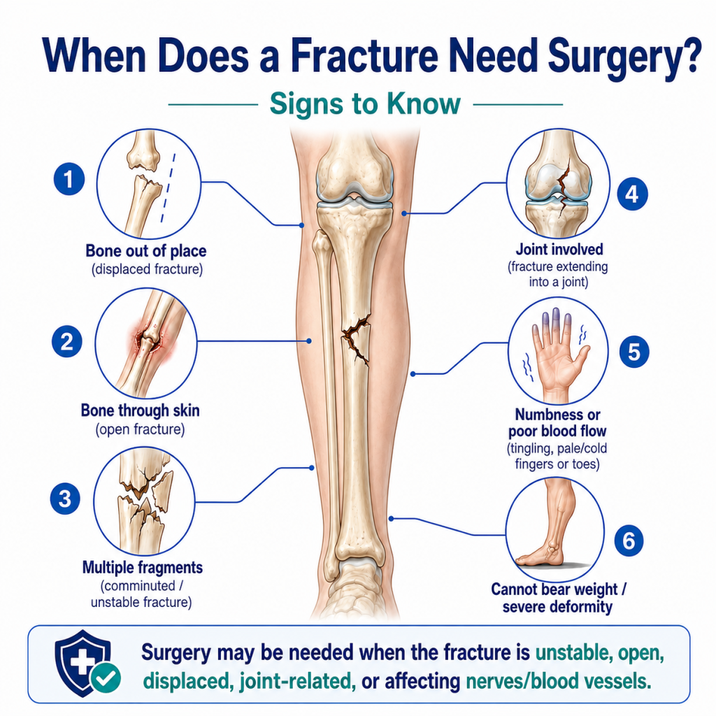
    A fracture needs surgery when the broken bone is badly displaced, unstable, open, involving a joint, affecting blood supply or nerves, not healing properly, or unlikely to stay aligned with only a cast or splint. Some fractures heal well with plaster, brace, or non-surgical treatment, but others require surgical fixation with plates, screws, rods, pins, or external support to restore alignment and function.

    At Shirdi Sai Hospital, New BEL Road, fracture treatment is planned after clinical examination and imaging. The goal is not to operate on every fracture. The goal is to choose the safest treatment that helps the bone heal in the correct position and allows the patient to regain movement, strength, and daily function.

    Medical infographic showing six warning signs that a fracture may need surgery, including displaced bone, open fracture, multiple fragments, joint involvement, numbness or poor blood flow, and inability to bear weight.

    What Is a Fracture?

    A fracture means a broken bone. The break may be small, partial, complete, displaced, open, or broken into multiple pieces. The severity depends on the force of injury, the bone involved, the patient’s age, bone strength, and whether nearby joints, nerves, blood vessels, skin, or muscles are affected.

    Common causes of fractures include:

    • falls
    • road traffic accidents
    • sports injuries
    • direct blows
    • twisting injuries
    • osteoporosis-related weak bones
    • repetitive stress injuries
    • workplace injuries

    Not every fracture looks dramatic. Some patients can still move the injured part, even with a fracture. That is why pain, swelling, deformity, inability to bear weight, or difficulty using the limb should be evaluated early.

    Does Every Fracture Need Surgery?

    No. Many fractures do not need surgery. If the bone pieces are well aligned, stable, and expected to heal properly, treatment may include a splint, plaster cast, brace, rest, medicines, and follow-up X-rays.

    Non-surgical fracture treatment may be suitable when:

    • the broken bone is not displaced
    • the fracture is stable
    • joint alignment is not affected
    • the bone can heal safely in a cast or brace
    • there is no open wound
    • there is no nerve or blood vessel injury
    • the patient can follow immobilization and follow-up advice

    However, some fractures cannot heal correctly unless the bones are realigned and stabilized surgically. This decision should be made by an orthopedic specialist after reviewing the X-ray or scan.

    When Does a Fracture Need Surgery?

    Fracture surgery may be needed when the bone position, stability, or injury severity makes non-surgical healing unsafe or unreliable.

    Common reasons include:

    • displaced fracture
    • open fracture
    • unstable fracture
    • fracture involving a joint
    • fracture with multiple bone fragments
    • fracture with nerve or blood vessel injury
    • fracture that cannot be held in a cast
    • fracture with severe soft tissue injury
    • hip fracture in elderly patients
    • fracture that is not healing properly
    • fracture that healed in the wrong position
    • repeated loss of alignment on follow-up X-rays

    Surgery is not chosen only because the fracture is painful. Pain is common in fractures. Surgery is considered when alignment, stability, healing, or function is at risk.

    Signs That a Broken Bone May Need Surgery

    A fracture may need surgical evaluation if the injury shows warning signs.

    Important signs include:

    • visible deformity
    • bone seen through the skin
    • severe swelling
    • severe pain after injury
    • inability to move the limb
    • inability to stand or walk after injury
    • numbness or tingling
    • fingers or toes becoming cold, pale, or blue
    • wound near the fracture site
    • severe pain with tight swelling
    • repeated fracture displacement
    • fracture near a joint
    • fracture after high-energy trauma

    These symptoms do not automatically mean surgery is certain, but they do mean urgent orthopedic evaluation is important.

    Fracture Surgery vs Cast: Main Difference

    FactorCast or Splint TreatmentFracture Surgery
    Used whenFracture is stable and well alignedFracture is unstable, displaced, open, or complex
    Main purposeImmobilizes bone externallyRealigns and stabilizes bone internally or externally
    Tools usedPlaster, splint, bracePlates, screws, rods, pins, external fixator
    Hospital stayOften shorter or outpatientDepends on fracture and procedure
    Follow-upX-rays to confirm alignment and healingX-rays, wound care, implant and rehab follow-up
    RecoveryDepends on fracture typeDepends on surgery, bone healing, and rehabilitation
    Risk if wrong choiceMalalignment, stiffness, delayed healingSurgical risks if not medically suitable

    This comparison helps patients understand why treatment choice is based on fracture stability, not only pain level.

    Types of Fractures That Often Need Surgery

    Some fracture patterns are more likely to need surgical management.

    Open Fractures

    An open fracture means the broken bone is associated with a wound in the skin. This is serious because infection risk is higher. Open fractures often need urgent cleaning, antibiotics, stabilization, and surgical care.

    Warning signs include:

    • wound near the broken bone
    • bone visible through skin
    • heavy bleeding
    • severe contamination with mud or road debris
    • severe pain and swelling after trauma

    Open fractures should be treated as urgent.

    Displaced Fractures

    A displaced fracture means the bone pieces have moved out of their normal alignment. If the bones cannot be aligned and held with a cast, surgery may be needed.

    Possible signs include:

    • limb looks bent or abnormal
    • shortening of the injured limb
    • severe swelling
    • abnormal movement
    • pain with deformity

    Proper alignment is important because bones that heal in the wrong position can affect movement, strength, and function.

    Unstable Fractures

    An unstable fracture may move even after being placed in a cast. If the bone cannot stay in the correct position, surgery may be advised to hold it securely.

    Examples include:

    • fractures with multiple fragments
    • certain long bone fractures
    • fractures with ligament injury
    • fractures that shift on follow-up X-rays

    Joint Fractures

    Fractures that extend into a joint may require surgery if the joint surface is not aligned properly. Joint alignment matters because uneven healing can lead to stiffness, pain, and early arthritis.

    Common joint areas include:

    • ankle
    • wrist
    • elbow
    • knee
    • shoulder
    • hip

    A fracture near a joint should be assessed carefully with imaging.

    Hip Fractures

    Hip fractures, especially in elderly patients, often need surgical management because long bed rest can lead to complications such as weakness, chest infection, clots, pressure sores, and loss of mobility.

    Treatment depends on fracture type, age, bone quality, mobility level, and medical condition.

    Fractures With Nerve or Blood Vessel Injury

    If a fracture affects nerves or blood vessels, urgent care is required. Signs may include:

    • numbness
    • tingling
    • weakness
    • loss of movement
    • cold fingers or toes
    • pale or bluish skin
    • absent pulse in the limb

    These symptoms need immediate medical attention.

    Fractures That Do Not Heal Properly

    Some fractures heal slowly or fail to heal. This is called delayed union or non-union. Surgery may be considered when healing is not progressing properly.

    Risk factors include:

    • smoking
    • diabetes
    • poor blood supply
    • infection
    • severe fracture
    • poor immobilization
    • repeated movement at fracture site
    • poor nutrition
    • certain medical conditions

    What Types of Bone Breaks Usually Require Surgery?

    The need for surgery depends on the exact fracture, but the following types commonly need orthopedic surgical evaluation:

    • open fractures
    • severely displaced fractures
    • comminuted fractures
    • unstable fractures
    • joint surface fractures
    • hip fractures
    • fractures with dislocation
    • fractures with nerve or blood vessel injury
    • some ankle fractures
    • some wrist fractures
    • some forearm fractures in adults
    • some thigh bone and leg bone fractures
    • fractures that fail to heal
    • fractures that heal in poor alignment

    The final decision depends on X-ray findings, patient health, age, function needs, and surgeon assessment.

    How Orthopedic Doctors Decide Whether Surgery Is Needed

    A fracture treatment plan is based on clinical examination and imaging.

    The orthopedic doctor checks:

    • which bone is broken
    • where the fracture is located
    • whether the bone pieces are aligned
    • whether the fracture enters a joint
    • whether the fracture is stable
    • whether the skin is open
    • whether nerves or blood vessels are affected
    • the patient’s age
    • activity level
    • bone quality
    • diabetes, blood pressure, or other medical conditions
    • whether the patient can safely undergo surgery
    • whether the fracture can be held with a cast

    In some cases, the doctor may advise a cast first and review healing with repeat X-rays. In other cases, early surgery may be safer.

    Tests Used Before Fracture Surgery

    The required tests depend on the fracture and patient condition.

    Common evaluations may include:

    • X-ray
    • CT scan for complex fractures or joint fractures
    • blood tests
    • ECG when needed
    • anesthesia fitness assessment
    • blood sugar and blood pressure review
    • infection evaluation in open wounds
    • neurovascular assessment
    • pre-surgery medical clearance in elderly or high-risk patients

    Imaging helps doctors understand the fracture pattern. Pre-surgery evaluation helps reduce avoidable risk.

    What Happens During Fracture Surgery?

    Fracture surgery aims to bring the broken bone pieces into proper alignment and hold them stable while the bone heals.

    Depending on the fracture, the surgeon may use:

    • plates and screws
    • rods or nails
    • pins or wires
    • external fixator
    • joint-specific implants
    • bone graft in selected cases

    The common surgical principle is reduction and fixation. Reduction means correcting the position of the bone. Fixation means holding it stable.

    Not every patient needs the same implant. The choice depends on fracture type, bone involved, age, bone quality, soft tissue condition, and recovery plan.

    What Is ORIF in Fracture Surgery?

    ORIF means Open Reduction and Internal Fixation.

    • Open reduction means the surgeon exposes the fracture area and aligns the bone.
    • Internal fixation means plates, screws, rods, or pins are used to hold the bone.

    ORIF may be used in displaced, unstable, complex, or joint-related fractures when accurate alignment is important.

    What Is External Fixation?

    External fixation uses pins or screws placed into bone above and below the fracture. These are connected to an external frame outside the body.

    It may be used when:

    • there is severe soft tissue injury
    • swelling is significant
    • the fracture is open
    • temporary stabilization is needed before another surgery
    • the fracture pattern needs external support

    External fixation is especially useful when the skin, muscles, or wound around the fracture need ongoing care.

    Recovery After Fracture Surgery

    Recovery depends on fracture type, surgery type, age, bone health, rehabilitation, and associated injuries.

    Recovery may include:

    • pain control
    • wound care
    • medicines as prescribed
    • limb elevation
    • movement restrictions
    • follow-up X-rays
    • physiotherapy
    • gradual weight bearing
    • implant monitoring
    • return-to-work guidance
    • follow-up for stiffness or weakness

    Pain often improves before the bone is strong enough for full activity. Patients should not return to lifting, running, sports, or heavy work without doctor clearance.

    Fracture Healing: What Patients Should Know

    Fracture healing is a biological process. Surgery helps align and stabilize the bone, but the body still needs time to heal.

    Healing depends on:

    • type of fracture
    • blood supply
    • age
    • nutrition
    • diabetes control
    • smoking status
    • infection risk
    • bone quality
    • follow-up compliance
    • physiotherapy and movement plan

    Healthy nutrition, doctor-guided activity, and follow-up care support recovery.

    What Can Go Wrong If Surgery Is Delayed?

    Delaying care for a fracture that needs surgery can increase risk.

    Possible problems include:

    • bone healing in wrong position
    • non-union
    • delayed union
    • joint stiffness
    • chronic pain
    • deformity
    • nerve or blood vessel damage worsening
    • infection in open fractures
    • poor function
    • more complex surgery later

    Not every fracture is urgent, but fractures with deformity, open wound, severe swelling, numbness, reduced circulation, fever, or inability to bear weight need timely evaluation.

    How to Choose a Qualified Orthopedic Surgeon

    Patients often search for how to choose an orthopedic surgeon for fracture treatment. The right surgeon should evaluate the fracture carefully and explain the treatment options clearly.

    Look for:

    • orthopedic training and experience
    • ability to explain X-ray findings
    • clear discussion of surgery vs non-surgery options
    • structured follow-up plan
    • rehabilitation guidance
    • patient safety focus
    • emergency support if needed
    • hospital facility for imaging and surgery

    Avoid choosing treatment based only on fear, cost, or online opinions. Fracture care should be based on diagnosis.

    Why Patients Choose Shirdi Sai Hospital for Fracture Care

    At Shirdi Sai Hospital, New BEL Road, fracture care is evaluated through the Orthopedics department with attention to the type of fracture, alignment, stability, swelling, pain level, and patient safety. The hospital’s Orthopedics department supports diagnosis and treatment of fractures, joint conditions, sports injuries, musculoskeletal problems, and rehabilitation needs.

    Patients may choose Shirdi Sai Hospital because:

    • Orthopedics services are available on New BEL Road
    • fracture care includes evaluation, imaging guidance, and treatment planning
    • surgical and non-surgical options are considered based on fracture type
    • emergency care is available for sudden injuries
    • rehabilitation guidance supports recovery and mobility
    • patients can be assessed for associated injuries after falls or accidents
    • the hospital is accessible for patients from Devasandra, Mathikere, RMV 2nd Stage, Sanjaynagar, Hebbal, and nearby areas

    If you are unsure whether your fracture needs a cast, brace, or surgery, you can consult the Orthopedics department at Shirdi Sai Hospital.

    When to Visit the Hospital Immediately

    Go to the hospital immediately if there is:

    • bone visible through skin
    • severe deformity
    • severe swelling
    • numbness or tingling
    • cold or blue fingers or toes
    • heavy bleeding
    • severe pain after injury
    • inability to stand or walk
    • suspected hip fracture
    • injury after road traffic accident
    • fracture with fever or wound infection
    • child with severe limb deformity
    • elderly person unable to move after fall

    If the injury is severe, do not massage, force movement, or try to “set” the bone at home.

    First Aid Before Reaching the Hospital

    Until medical care is available:

    • keep the injured part still
    • avoid putting weight on the injured limb
    • apply a clean cloth over any open wound
    • use a splint only if trained or if safe support is available
    • apply cold pack wrapped in cloth for swelling
    • keep the limb elevated if possible
    • do not push visible bone back inside
    • do not massage the injured area
    • do not eat or drink heavily if surgery may be needed

    Emergency evaluation is the priority in severe injuries.

    Frequently Asked Questions

    When does a fracture need surgery?

    A fracture may need surgery when the bone is displaced, unstable, open, involving a joint, affecting nerves or blood vessels, broken into multiple pieces, or unlikely to stay aligned with a cast.

    What are the signs that a broken bone needs surgery?

    Warning signs include visible deformity, open wound, severe swelling, numbness, cold fingers or toes, inability to bear weight, severe pain, or a fracture near a joint.

    Can all fractures heal without surgery?

    No. Many stable fractures heal with cast or splint treatment, but unstable, displaced, open, joint-related, or complex fractures may need surgery.

    What types of fractures usually require surgery?

    Open fractures, displaced fractures, unstable fractures, comminuted fractures, joint fractures, hip fractures, fractures with dislocation, and fractures with nerve or blood vessel injury often need surgical evaluation.

    Is fracture surgery risky?

    Every surgery has risks such as infection, bleeding, stiffness, anesthesia risk, and delayed healing. However, when surgery is required, delaying it may cause worse alignment, poor function, or complications.

    How do doctors decide if fracture surgery is needed?

    Doctors assess the X-ray or scan, fracture location, alignment, stability, soft tissue injury, nerve and blood vessel status, patient age, bone quality, and overall health before deciding treatment.

    Does pain mean a fracture needs surgery?

    No. Pain is common in fractures. Surgery is considered when alignment, stability, joint involvement, circulation, nerve function, or healing is at risk.

    Conclusion

    A fracture does not always need surgery. Many stable fractures heal with a cast, splint, or brace. Surgery becomes important when the bone is displaced, unstable, open, near a joint, affecting blood vessels or nerves, or unlikely to heal correctly without fixation.

    At Shirdi Sai Hospital, New BEL Road, fracture treatment is planned after proper orthopedic evaluation and imaging. If you have a suspected fracture, severe swelling, deformity, open wound, numbness, or inability to bear weight, timely consultation can help prevent complications and support better recovery.

    OPD Consultation starts from ₹500.