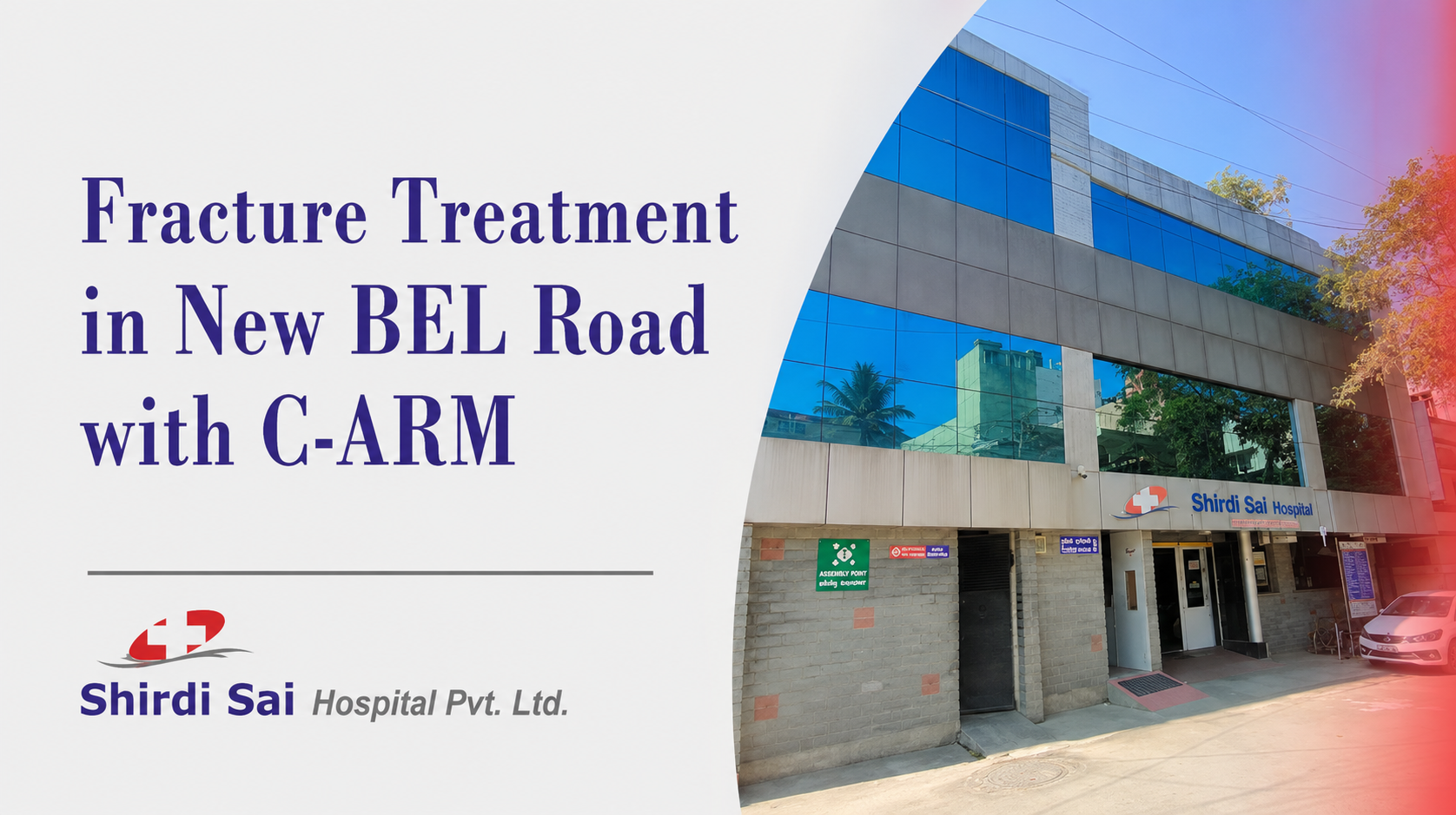
Shirdi Sai Hospital provides fracture treatment in New BEL Road for patients with suspected broken bones, pain after a fall, swelling, deformity, difficulty moving a limb, or inability to bear weight. Care begins with an orthopaedic assessment and appropriate imaging to understand which bone is injured, whether the fracture is stable, and whether nearby joints, nerves, blood vessels, or soft tissues are affected. Some fractures may be treated with a splint, cast, or brace. Others may require realignment or surgical fixation. C-Arm imaging is available for selected orthopaedic procedures when real-time X-ray guidance is clinically useful.
What Is a Bone Fracture?
A fracture is a partial or complete break in a bone. The words “fracture” and “broken bone” describe the same type of injury. A fracture may occur after a fall, road accident, sports injury, direct blow, or repeated stress. Weakened bones, including bones affected by osteoporosis, may sometimes fracture after a relatively minor injury.
Common fracture patterns include:
- Stable or non-displaced fracture: The broken ends remain reasonably aligned.
- Displaced fracture: The bone fragments have moved out of their normal position.
- Open fracture: A wound communicates with the fracture site, increasing the need for urgent care.
- Comminuted fracture: The bone has broken into several pieces.
- Stress fracture: A small crack develops after repeated loading or overuse.
- Joint-involving fracture: The break extends into a joint surface and may affect movement.
- Pathological fracture: A weakened bone breaks because of an underlying condition.
The fracture pattern, location, alignment, skin condition, and effect on nearby tissues all influence treatment.
How Do You Know Whether a Bone Is Broken?
A fracture cannot always be confirmed from symptoms alone. Severe sprains, dislocations, and soft-tissue injuries can cause similar pain and swelling.
Possible fracture symptoms include:
- Pain after a fall, accident, or direct impact
- Swelling and tenderness
- Bruising
- Difficulty moving the injured area
- Pain while standing or bearing weight
- A limb or joint that looks bent or out of position
- A grinding or cracking sensation during the injury
- Numbness or tingling beyond the injured area
- An open wound near the suspected fracture
- Refusal to use an injured arm or leg in a child
An X-ray is commonly used to confirm whether a bone is broken and assess its position. CT or other imaging may be recommended for selected complex fractures or when the injury involves a joint.
What Is C-Arm Imaging in Fracture Treatment?
A C-Arm is a movable fluoroscopic X-ray system that provides real-time images during selected procedures. The C-shaped equipment can be positioned around the patient to produce images from different angles. It may help the treating surgeon view bone alignment, fracture reduction, surgical instruments, and implant position during a procedure.
At Shirdi Sai Hospital, C-Arm assistance is listed as available for selected orthopaedic procedures when the treating team considers it clinically appropriate. It may be used during certain fracture fixations, implant placements, trauma procedures, or other operations where live bone imaging would help guide the procedure.
What C-Arm guidance may help the surgeon assess
During an appropriate fracture procedure, C-Arm images may help with:
- Viewing the fracture from different angles
- Checking whether bone fragments have been realigned
- Guiding the placement of wires, screws, plates, or rods
- Confirming implant position during the procedure
- Reviewing alignment before the procedure is completed
- Supporting selected procedures performed through smaller operative access
C-Arm is an imaging aid. It does not replace the surgeon’s assessment, clinical judgement, or preoperative planning.
Does Every Fracture Need C-Arm or Surgery?
No. Many fractures may heal after proper realignment and immobilisation with a splint, cast, or brace. Surgery is more likely to be considered when a fracture is displaced, unstable, open, fragmented, extends into a joint, or cannot be kept in a suitable position using non-surgical treatment.
C-Arm imaging is generally relevant only when real-time guidance would help during reduction or surgical treatment. It is not normally required for every fracture consultation, routine cast application, or uncomplicated injury.
The decision depends on:
- Which bone is broken
- The fracture pattern
- Whether the fragments have moved
- Whether a joint is involved
- The patient’s age
- Bone quality
- Skin and soft-tissue injuries
- Nerve and blood-vessel function
- Other medical conditions
- The patient’s mobility and functional needs
A patient should not select surgery or request a specific implant based only on an online description. The appropriate treatment requires examination and review of current imaging.
Fracture Evaluation at Shirdi Sai Hospital
Initial injury assessment
The medical team may ask:
- How the injury occurred
- When it happened
- Where the pain is located
- Whether the patient can move the limb
- Whether weight can be placed on the injured side
- Whether numbness or weakness is present
- Whether there is bleeding or an open wound
- Whether the patient struck their head
- Whether there are other injuries
- Which medicines the patient takes
- Whether the patient has diabetes, osteoporosis, or another medical condition
Physical examination
The injured area may be examined for:
- Swelling and tenderness
- Bruising
- Visible deformity
- Skin wounds
- Joint movement
- Sensation
- Muscle movement
- Pulse and blood circulation beyond the injury
- Signs of damage to nearby nerves or blood vessels
Assessment of circulation, sensation, skin condition, and associated injuries is important because a fracture can affect more than the bone itself.
Imaging
The investigation plan may include:
- X-ray: Commonly used to identify a fracture and assess alignment.
- CT scan: May provide additional detail for complex or joint-involving fractures.
- MRI: May be considered when an occult fracture or associated soft-tissue injury is suspected.
- Repeat imaging: Sometimes required during follow-up to check whether the position remains suitable and healing is progressing.
Not every fracture requires advanced imaging.
Fracture Treatment Options
The appropriate treatment depends on the stability and position of the fracture, the affected bone, the surrounding tissues, and the patient’s overall condition.
Splinting
A splint may be applied soon after an injury to restrict movement and support the limb while allowing space for swelling. It may be used temporarily before a full cast or before a treatment decision is finalised.
Casting or bracing
A cast or brace may be suitable when the fracture is stable or can be maintained in an acceptable position without surgery. Follow-up imaging may be needed because some fractures can shift while swelling settles or healing begins.
Closed reduction
Closed reduction means repositioning displaced bone fragments without surgically opening the fracture site. After realignment, the injured area may be supported with a cast, splint, brace, pins, or another stabilisation method.
Pain control, sedation, anaesthesia, and imaging support depend on the procedure and patient.
Surgical fracture fixation
Surgery may be considered when adequate alignment or stability cannot be achieved with a cast or brace.
Possible fixation methods include:
- Plates and screws
- Intramedullary rods or nails
- Pins or wires
- External fixation
- Other implants selected for the fracture pattern
During open reduction and internal fixation, the bone fragments are repositioned and held using suitable implants. External fixation uses pins or screws connected to a frame outside the body and may be considered when the fracture or surrounding soft tissues require that approach.
Wound and soft-tissue care
An open fracture requires urgent evaluation because the broken skin creates a risk of contamination and infection. Treatment may involve wound cleaning, antibiotics, fracture stabilisation, and one or more procedures depending on the injury.
Physiotherapy and rehabilitation
Rehabilitation may begin at different stages depending on the fracture and its stability. The goals may include restoring joint movement, muscle strength, balance, walking ability, and safe return to daily activity.
Exercises should follow the treating team’s guidance. Starting activity too early or placing weight on the injured limb before approval may affect healing.
Fractures That May Require Orthopaedic Care
The Orthopaedics department may evaluate fractures involving:
- Wrist and forearm
- Hand and fingers
- Elbow
- Upper arm
- Collarbone and shoulder
- Ribs
- Hip and pelvis
- Thigh bone
- Knee region
- Leg
- Ankle and foot
- Spine
- Bones affected by osteoporosis
- Sports-related stress fractures
- Fractures in children and older adults
Treatment differs considerably between these injuries. A stable finger fracture does not follow the same pathway as a hip fracture, open leg fracture, or joint-involving wrist fracture.
When Is a Fracture an Emergency?
Seek urgent hospital assessment after an injury when there is:
- A visibly bent or deformed limb
- Bone visible through a wound
- A deep wound over the injured area
- Heavy or uncontrolled bleeding
- Severe or increasing pain
- Inability to stand or bear weight
- Numbness or loss of sensation
- Fingers or toes that appear pale, blue, or unusually cold
- Severe swelling or a very tight feeling in the limb
- Suspected hip fracture after a fall
- Suspected spine injury
- Head injury, fainting, confusion, or vomiting after an accident
- Multiple injuries following a road accident or fall from height
- A child refusing to move an injured limb
Open fractures, significant deformity, circulation changes, and severe trauma require prompt assessment. Shirdi Sai Hospital currently lists 24-hour emergency care for injuries, trauma, and suspected fractures at its New BEL Road facility.
What Should You Do Before Reaching the Hospital?
While arranging medical care:
- Keep the injured area as still as reasonably possible.
- Avoid walking on a painful or visibly injured leg.
- Support the limb using soft padding when safe to do so.
- Apply gentle pressure with a clean cloth if there is bleeding.
- Cover an open wound with a clean dressing.
- Do not attempt to push an exposed bone back into the wound.
- Do not repeatedly test the movement of the injured limb.
- Avoid unnecessary movement when a spine, neck, or major trauma injury is suspected.
- Seek safe transport to an appropriate emergency department.
Do not delay urgent care to collect reports or wait for swelling to settle.
What Happens After Fracture Treatment?
Follow-up is important even when the initial treatment is non-surgical.
The care plan may include:
- Repeat X-rays
- Cast, splint, or wound review
- Monitoring of swelling and circulation
- Suture or dressing care after surgery
- Medication guidance
- Instructions about weight bearing
- Implant review
- Physiotherapy
- Gradual return to work or sport
- Evaluation for osteoporosis in relevant patients
Healing time cannot be predicted from the fracture name alone. It varies with the affected bone, fracture severity, age, blood supply, general health, treatment stability, smoking status, nutrition, and adherence to medical instructions. Pain may improve before the bone is ready for normal loading, so activity should be resumed only according to clinical guidance.
Warning Signs During Recovery
Contact the treating team or seek urgent assessment if there is:
- Increasing pain that is not settling
- New numbness or burning
- Fingers or toes becoming pale, blue, cold, or increasingly swollen
- A cast that feels excessively tight
- Fever or chills
- Fluid, bad smell, or increasing discharge from a wound or cast
- Sudden loss of movement
- A new injury to the treated area
- Increasing redness around a surgical wound
- Breathlessness or chest pain during recovery
A cast should not be modified or removed at home unless the treating team has given specific instructions.
Fracture Treatment in New BEL Road
Shirdi Sai Hospital is located at No. 519, 2nd Main, Nethravathi Street, Devasandra, New BEL Road, Bengaluru, Karnataka 560054. Its live Orthopaedics content lists fracture evaluation, casting, bracing, surgical treatment when required, rehabilitation guidance, and C-Arm assisted orthopaedic procedures where clinically indicated.
Patients may approach the hospital for:
- Assessment after a fall or accident
- Suspected fracture
- Review of an existing X-ray or scan
- Splint or cast evaluation
- Fracture realignment assessment
- Surgical fracture-fixation planning
- C-Arm assisted procedures where indicated
- Follow-up after fracture treatment
- Rehabilitation guidance
- A second orthopaedic opinion
The suitability of a procedure, implant, or admission pathway depends on clinical evaluation. Current orthopaedic doctor and procedure availability should be confirmed directly before a planned visit.
Confirm the Correct Shirdi Sai Hospital
Shirdi Sai Hospital is a separate organisation from hospitals using the name Sathya Sai Hospital.
Before requesting directions, confirm:
Hospital: Shirdi Sai Hospital Pvt. Ltd.
Location: Devasandra, New BEL Road, Bengaluru 560054
Phone: +91 8088511289
Official website: Shirdi Sai Hospital Pvt. Ltd.
The live contact page confirms the New BEL Road address and official phone number.
Book a Fracture Consultation in New BEL Road
For a planned orthopaedic consultation, call +91 8088511289 and briefly explain:
- How the injury occurred
- Which body part is affected
- When the injury happened
- Whether the patient can move or bear weight
- Whether an X-ray has already been taken
- Whether there is a wound, deformity, or numbness
For a suspected fracture with severe pain, deformity, an open wound, inability to walk, circulation changes, or a significant accident, seek emergency assessment rather than waiting for a routine appointment or website response. The hospital’s contact details and emergency pathway are currently live.
Frequently Asked Questions
Does every fracture require surgery?
No. Stable fractures may sometimes be treated with a splint, cast, or brace. Surgery may be considered when the fracture is displaced, unstable, open, extends into a joint, or cannot be maintained in a suitable position without fixation.
What is C-Arm used for during fracture treatment?
C-Arm is a movable fluoroscopic X-ray system that provides real-time images during selected procedures. It may help the surgeon assess fracture alignment and guide the placement of implants such as screws, plates, pins, or rods.
Is C-Arm needed for every fracture?
No. C-Arm is used only when live imaging is considered useful during reduction or surgical treatment. Routine consultations, uncomplicated casting, and many stable fractures may not require it.
How is a fracture diagnosed?
The doctor examines the injured area and checks movement, swelling, skin condition, circulation, and sensation. An X-ray is commonly used to confirm the fracture. CT or other imaging may be advised for selected complex injuries.
When should I go to emergency care for a suspected fracture?
Seek urgent care for visible deformity, an open wound, bone protruding through the skin, severe pain, inability to bear weight, numbness, uncontrolled bleeding, or fingers or toes becoming pale, blue, or cold.
How long does a fracture take to heal?
Healing time varies according to the bone involved, fracture pattern, age, treatment, blood supply, and medical condition. Some fractures heal within several weeks, while others can take several months. The treating orthopaedic doctor should guide activity and follow-up based on imaging and progress.
How can I arrange fracture treatment in New BEL Road?
Call Shirdi Sai Hospital on +91 8088511289 and ask for the Orthopaedics or emergency pathway based on symptom severity. Confirm appointment and doctor availability before a planned visit. For a serious injury or visible deformity, seek urgent assessment.